
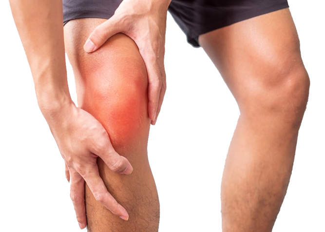
Nerve Injuries Leg
Nerve Injuries Leg
Nerves are the "electrical wiring" system of the body that carries messages between the brain and the rest of the body.
Nerves are the "electrical wiring" system of the body that carries messages between the brain and the rest of the body. There are two types of nerves- motor and sensory nerves. Motor nerves carry messages between the brain and muscles to make the body move. Sensory nerves carry messages between the brain and different parts of the body to signal pain, touch and temperature.
The nerves can malfunction if compressed, stretched or injured.
When the nerve is compressed or stretched, outer cover of the nerve remains intact, but the ability of the nerve to send and receive signals from various parts of the body is compromised. If this compression is unrelieved, it can cause permanent damage to the nerve
When the nerve is cut or injured, fascicles within the nerve and its outer cover both are damaged with partial or complete loss of motor power and sensory input from the area of the distribution of nerve. If the nerve is not repaired, the growing nerve fibers may form a painful nerve scar, or neuroma at the cut ends.
Think of nerve as an electric wire And injury to nerve as a cut electric wire. The cut ends of the electric wire should be brought and glued together so that the electricity can travel through the wire uninterrupted and reach its destination like fan, tubelight. Similarly, cut ends of the nerve should be brought together surgically and sutured under microscope with very fine sutures so that signals from the brain can reach to different parts of the body.
But there is a big difference!!! There is immediate flow of electricity through the repaired wire in contrast to the nerve repair which takes a much longer time. Nerve fibers typically begin to regrow about three or four weeks after surgery. The nerve grows at a constant speed of one inch per month after it is repaired. So, the results of nerve repair can take months to a year to appear. During the process of recovery, feeling of pins and needles in area of distribution of the nerve is common. While this can be uncomfortable, it usually passes and is a sign of recovering nerve.
Timing of Nerve Repair
After nerve repair, new fibers grow beneath the covering layer until it reaches a muscle or sensory receptor. The nerve fibers should reach the target muscle within 18 months of the injury otherwise the muscle receptor degenerates. For this reason, repair should be done as early as possible after injury to allow maximum time period for growth of the nerve.
The sensory receptor does not degenerate, therefore, sensory nerve repair can be done anytime or years after the injury also.
Management of Nerve Injury
- If the nerve is compressed or stretched, removal of the offending structure or situation will help in spontaneous healing and recovery of the nerve.
- If the nerve is cut and wound is clean, covering around both the cut ends of the injured nerve is sewn together. The goal in repairing the nerve is to suture the covering layer with minute sutures so that new fibers can grow forward across the injured site towards its target organ and the nerve can work again.
- If a nerve is cut and wound is dirty or crushed, surgery may be delayed until the skin has healed.
- If there is a space between the ends of the nerve, it may be necessary to take a piece of nerve (nerve graft) from another part of the body to repair the injured nerve.
- The results of nerve repair depends on a number of factors like age of the patient, presence of other diseases and the level of injury etc.
- Nerve fibers typically begin to regrow about three or four weeks after surgery. During this time, patients must wear a splint to prevent the repaired nerve from stretching apart.
- Physiotherapy is must after nerve repair to keep the joints supple
- Care must be taken to prevent oneself from burn or other injuries till the sensation returns back in cases of sensory nerve repair.

Nerve repair with realignment of bundles.
Sciatic Nerve Injuries
Sciatic nerve branches from the lower back region passes through the gluteal region, thigh and divides into 2 main branches in the lower part of the thigh-common peroneal nerve and tibial nerve.

Sciatic nerve supplies muscles of the back of thigh and apparently to all the muscle of whole of leg and foot viaits two main branches named above. Sciatic nerve is responsible for sensation in the whole of the leg and foot.
Sciatic nerve can be injured by accidents, compression or by stretching of the nerve. It can happen in fractures and dislocation of the hip. It is quite commonly wrongly injected with drugs and damaged while giving intramuscular injections by untrained professionals.
Injuries to the sciatic nerve can cause
- Inability to move the foot and toes- upward or downward
- Numbness on whole leg and foot.
- Tingling and pain on the leg and foot region.
- Loss of ability to move the foot forward due to weakness of the muscles of foot and leg.
- Tinel's sign will be positive– Tapping on the site of injury will elicit tingling and pain sensation in the leg and foot region.
- To confirm the diagnosis- Nerve Conduction Velocities (NCV) and Electro-Myographic Studies (EMG)of the affected limb is done.
- In few cases, CT scan or MRI is also indicated.
Sciatic nerve injury is a very debilitating condition, mainly because of 2 major deficiencies-
- complete loss of muscle power in foot and toes
- loss of sensation on sole of the foot -making patient prone to more injuries and burns over foot
Treatment of Sciatic Nerve Injury
- If the sciatic nerve is damaged or cut, then surgical excision of the damaged part and nerve repair/grafting should be done as early as possible or within 3 months of the injury. The results of repair are not good for sciatic nerve injuries as the injury is most of the times so up and proximal that by the time nerve fibers grow and reach up to the leg region, motor end plates or receptors had already degenerated.
- If the patient has presented with sudden onset of paralysis of sciatic nerve, then other neurological causes or compression by tumor should be investigated for. In many of such cases, release of the nerve is done and a biopsy of the nerve fascicle and muscle is taken to confirm the diagnosis of neurological disease.
If the problem is caused by an underlying illness or some other fracture or dislocation or tumor, it is important to address that issue. - If the patient presents with gradual paralysis of sciatic nerve, then sciatic nerve compression - should be thought. Offending factor should be removed and release of the compression with creation of more space for the nerve should be done surgically.
- If the patient has presented late beyond the timing suitable for sciatic nerve repair (after 3 months to 6 months of the accident), then also nerve repair and grafting should be done. It might not help in restoration of the muscle function but can help in restoring the sensation to the sole of the foot. Tendon transfers for restoring the muscle movements of the foot cannot be done in sciatic nerve injuries as all the muscles are paralysed and no tendon is left for transfer.
The results of sciatic nerve repair is relatively good in children and in patients who come early. The result is better if direct nerve repair is possible without the use of nerve grafting and if the level of injury is not higher up and is in the lower thigh.
More than 50% of the patients require secondary procedures in form of microvascular free functioning transfer or arthrodesis of the foot even after nerve repair.
Free functioning muscle transfer can be attempted by microvascular surgical techniques to restore function of movement of the ankle and foot in upward direction. A strong muscle from the thigh is taken with its blood and nerve supply and fixed at 2 different points of the leg and foot. The blood and nerve supply of the muscle is joined with the blood and nerve supply in the region of leg by microsurgical techniques. The goal is to enable the patient to extend his or her foot and toes at their joints.
The surgery is usually done under spinal anaesthesia or general anaesthesia and a splint or slab is given for immobilization in the post operative period. The patient is usually discharged tenth day after the surgery. Sutures are removed3 weeks after surgery. Splint is removed 6 weeks after the surgery and gradual training of the transferred muscles is started so that they regain their newly assigned function.
Common Peroneal Nerve Injuries
Common peroneal nerve branches from the sciatic nerve in the lower part of the thigh and passes to the leg from its outer aspect. It then divides into 2 branches-, superficial and deep, which runs down the front of leg and foot region upto the tip of toes. This nerve provides sensation to the front and sides of the legs and to the top of the feet and toes. This nerve also controls the muscles in the leg that lift the ankle and toes upward. 
Common peroneal nerve can be injured by accidents, compression or by stretching of the nerve. It can happen in fractures and dislocation of the knee and leg.
Injuries to the peroneal nerve can cause
- Inability to point the toes upward or lift the ankle up (dorsiflexion)
- Numbness on the front of leg, foot and dorsum of toes
- Tingling and pain on the front of leg and foot region.
- Loss of ability to move the foot due to weakness of the muscles of foot and leg
- A distinctive style of walking is seen where the knee is raised higher than normal to clear the foot from the ground when the leg swings forward (also called step page or foot drop gait).
- Tinel's sign will be positive– Tapping on the site of injury will elicit tingling and pain sensation in the foot region.
- To confirm the diagnosis- Nerve Conduction Velocities (NCV) and Electro-Myographic Studies (EMG)of the affected leg is done
- In few cases, CT scan or MRI is also indicated
Treatment of Peroneal Nerve Injury
- If the peroneal nerve is damaged or cut, then surgical excision of the damaged part and nerve repair/grafting should be done as early as possible or within 6 months of the injury for best results.
- If the patient has presented with sudden onset of paralysis of peroneal nerve, then other neurological causes, leprosy or compression by tumor should be investigated for. In many of such cases, release of tunnel under which it passes on the outer aspect of the leg, is done and a biopsy of the nerve fascicle and muscle is taken to confirm the diagnosis of neurological disease or leprosy.
If the problem is caused by an underlying illness or some other fracture or dislocation or tumor, it is important to address that issue. - If the patient presents with gradual paralysis of peroneal nerve, then peroneal nerve compression - should be thought. Offending factor should be removed and release of the compression with creation of more space for the nerve should be done surgically
- If the patient has presented late beyond the timing suitable for peroneal nerve repair (after 6 months to 1 year of the accident) or in cases where nerve is damaged beyond repair, then tendon transfers should be done to strengthen the weak portions of the foot along with the nerve repair and grafting (only if feasible). A strong tendon of the leg is re-routed to substitute a weaker or lost movement of the foot.
During tendon transfers for peroneal nerve palsy, one functioning tendons from the inner side of the leg is taken, re-routed and sutured to the tendons of the front of foot and toes so that the patient can extend his or her foot and toes at their joints. This tendon transfer also enables and restores normal walking style.
The surgery is usually done under spinal anaesthesia or general anaesthesia and a splint or slab is given for immobilization in the post operative period. The patient is usually discharged second day after the surgery. Sutures are removed 14 days after surgery.
Splint is removed 4 weeks after the surgery and gradual training of the transferred muscles or tendons is started so that they regain their newly assigned function.
Shaping dreams through


Know your surgeon better
Best plastic surgeon, Dr. Amit Agarwal is an American Board Certified, extensively trained, and best Plastic & Aesthetic surgeon in Lucknow. He is the Chief Plastic Surgeon heading the Department of Plastic, Microvascular, and Craniofacial surgery at Vivekananda Polyclinic and Institute of Medical Sciences, Lucknow, U.P, India. He maintains a busy practice at Avadh and Nishat Hospital and his own center - Kayakriti Plastic Surgery & Dental Center. He was formerly a Consultant in the Department of Plastic Surgery and Burns at the prestigious SGPGI, Lucknow.
MS, DNB (General Surgery) MCh, DNB (Plastic Surgery),
MNAMS, FACS, FICS, FRCS (Edinburgh, UK)
His Credentials


Three pillars of kayakriti
Privacy
We believe your experience with us should be comfortable and hassle-free to make it one of your best lifetime experiences for yours. We, here at the clinic, take full precautions to maintain your privacy in any manner. We also provide a staff who will receive you from the gate and take you to the chamber directly if you demand.
Trust
Our Surgeon is highly qualified and internationally certified with a team of skilled staff to perform any surgical or non-surgical treatment on your body.
Safety
When you plan to undergo any surgery you should always keep in mind that it's your body and it's a surgery. We, here always keep your safety a priority and will never recommend you to undergo any such procedure which is not safe for you. We also provide you with a detailed description of the complications which may occur after the surgery during the consultation as it's a surgical procedure so there may be some complications depending on the way your body reacts.
Kayakriti in news


Frequently Asked Questions
If you have flat or small breast and you want to improve your breast and hip contour ratio then you are a good candidate for it. The answer will be best provided after the first consultation with Dr Amit Agarwal.
Acute pain will be there for almost a week which gradually reduces and there will be soreness and swelling which may take up to 3 weeks to subside.
You can join your work and daily routines after a week of the procedure and can start exercising after 3 weeks of it.
Yes, you have to wear it round the clock unless we suggest you to remove it.
This surgery does not affect the ducts or the areas of the breast involved in milk production. Thus, it does not affect the breast feeding.
This surgery does not affect the ducts or the areas of the breast involved in milk production. Thus, it does not affect the breast feeding.


Kayakriti Plastic Surgery & Dental Center
D-43, Near Punjab National Bank, Rajajipuram, Lucknow, Uttar Pradesh - 226017, India
Phone No. +919695940009, +919695940006
Map Location

Social Media Presence