LeFort Osteotomy
LeFort Osteotomy is the name of the surgical procedure and is usually indicated for the treatment of deformities of the middle of the face (bone named maxilla and zygoma).
Best LeFort Osteotomy Surgery in India
What is LeFort Osteotomy?
LeFort Osteotomy is the name of the surgical procedure and is usually indicated for the treatment of deformities of the middle of the face (bone named maxilla and zygoma). The upper jaw is surgically cut and repositioned to correct the deformity in this surgery
The common deformities corrected are
- Underdeveloped Upper Jaw (Maxilla).
- Overdeveloped Upper Jaw (Maxilla) resulting in a gummy smile
- Midface Skeletal Deformities.
What can you expect?
LeFort Osteotomy procedure is considered to be safe, reliable & has good long-term effects, and orthognathic surgery. Hence this procedure is being used very commonly. This procedure requires the incorporation of an Orthodontist for pre- & postoperative care for the correction of dentofacial deformities.
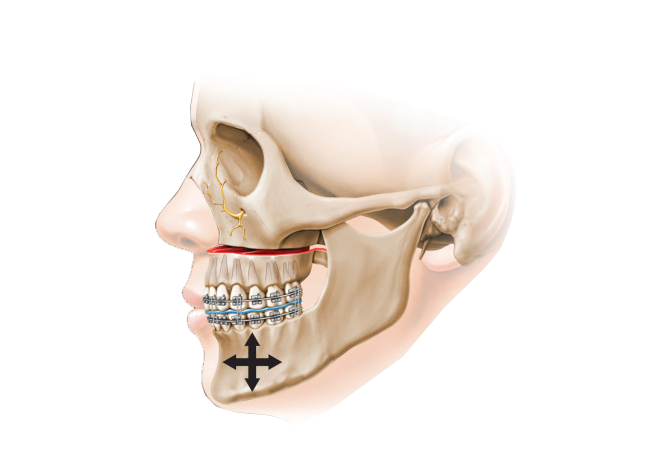
During this surgical procedure, the upper jaw is surgically separated from the facial bones. After separating the upper jaw, according to the facial deformity, it can be moved in either one or more of these directions-
Upward, downward, forward, backward, or tilted and turned After the upper jaw is moved to the desired location, it is fixed to the facial bones with the help of plates and screws
With the advent of the LeFort Osteotomy procedure, a complex range of mid-face deformities can be corrected. The reassembly of the entire facial complex is possible with the help of the LeFort Osteotomy procedure.
In severe deformities of the face, it can be combined with Bilateral Sagittal Split Osteotomy (BSSO). When Lef is combined with BSSO, it is called Bi Jaw/ Bi Max surgery.
Lefort Osteotomy is routinely combined with chin surgery/genioplasty.
Symptoms for LeFort Osteotomy Surgery
- Correction of malocclusion of teeth.
- Maxillomandibular (Jaw)Deformities.
- Facial Asymmetries.
- Underdeveloped maxilla in Clefts.
- Obstructive Sleep Apnea.
- Maxillary Atrophy.
When to get LeFort Osteotomy Surgery
- Before performing orthognathic surgery, it is important to check that the skeletal growth is complete in both boys and girls which is usually 15 to 16 years in girls and 17 to 18 in boys. Maxillary (Upper Jaw) growth usually ceases 2 years before Mandibular (Lower jaw) growth completion.
- Mandibular (Lower jaw) growth completion. The only contradiction to the above rule is -Surgical correction for maxillary excess during the growth period.
Pre operative planning at Kayakriti
Preoperative surgical planning is the key to successful orthognathic surgery
A) Investigations
- 3D CT scan of the face
- Frontal and lateral photos of the face
- Cephalometric X-Ray of the face for soft tissue and bony measurements.
- Dental impressions are obtained to aid in formulating a diagnosis.
Out of all the mentioned investigations, Cephalometric X-rays are very important as they reveal abnormal growth patterns of the upper and lower jaws about the cranial base.
B) Analysis
Then a detailed analysis of the skeletal, dental, and soft tissue findings is done by Dr. Amit Agarwal and the orthodontist. Dental models are made and a mock surgery is performed over the model and acrylic occlusal splints are fabricated. These occlusal splints are used intraoperatively to accurately reposition the osteotomized (cut) upper jaw - maxilla.
Contraindications for Lefort Osteotomy
- Major medical comorbidities
- Cardiac diseases
- Presence of active dental infections
- A patient in the active growth phase
- Psychiatric diseases or uncontrolled seizure disorders
- The major difference between patient's and surgeon's goals and expectations
- Patients with a habit of smoking and substance abuse
- Questionable blood supply of the maxilla as seen in multiply operated patients (e.g. cleft palate) or patients with a history of radiation therapy
10 Steps for Surgical Procedure of LeFort Osteotomy
- This procedure is performed under general anesthesia with nasal endotracheal intubation. Local anesthesia (lidocaine 1% with 1:100,000 epinephrine) is also used to decrease blood loss.
- During maxillary down fracture, the technique of hypotensive anesthesia will also lead to decreased blood loss.
- A vestibular intraoral incision is given to preserve the attached gingiva.
- The muscles attached to the bone are cut with a fine cautery and the periosteum which is the covering of the bone is reflected up.
- The important surgical bony landmarks are brought into view which includes the piriform nasal apertures, the lateral nasal walls, the nasal septum and vomer, the anterior nasal spine, the infraorbital foramen, and neurovascular bundle, and the lateral maxillary wall.
- The upper jaw (maxilla) is then cut and separated from the remaining facial bones and moved to its desired location which has been decided preoperatively.
- The most common separation is a Horizontal cut (Osteotomy) in which the surgeon uses the reciprocating saw to a cut (osteotomy) through the maxilla.
- A lot of care is taken to know the location of the apices of the tooth and the pterygoid plates posteriorly while cutting. Proper clinical examination & X-rays are required to prevent damage to the teeth due to the maxillary separation.
- If the gap between the bones is more than a centimeter and where a large gap exists between the segments after fixation, then bone grafting is done to prevent relapse and to provide long-term stability.
- Temporary inter-maxillary fixation with elastics is done over pre-surgically made occlusion wafers. The surgically moved maxilla is fixed with the native bones of the face with the help of plates and screws and the incision is closed in layers with absorbable sutures.
A) Le Fort Osteotomy in Cleft Patients
Le Fort 1 Osteotomy with horizontal advancement is done for cleft children after their skeletal growth is complete. It is the last stage of correction in cleft patients. As cleft patients mostly have a Class III malocclusion with narrow dental arch & palatal collapse, their facial height is also reduced.
Though Le Fort 1 Osteotomy with advancement is the standard method in many cases Segmental Le Fort 1 Osteotomy with Orthodontic Rapid Palatal Expansion has also been advised.
B) Le Fort Osteotomy in Class II Deformity
This deformity is mostly seen in cases of the underdeveloped or retruded mandible. In most of the cases, mandibular advancement & genioplasty is done but Le Fort 1 Osteotomy & repositioning is also advised in a few cases along with mandibular advancement.
C) Lefort Osteotomy in Gummy Smile Patients
Patients with an increased vertical height of the maxilla are also benefited from this surgery as this surgery helps to decrease the height of the upper jaw in the vertical direction which reduces the gingival show.
D) Lefort Osteotomy in Obstructive Sleep Apneoa (OSA)
Airway obstruction in case of Obstructive sleep apnea can also be well treated with this surgery. The airway is first properly evaluated with proper investigations, to determine the cause of OSA and to know if there is a skeletal obstruction for which advancement of the upper jaw can be done to increase the volume of the oro- & nasopharyngeal airway. This procedure in selected patients gives relief to them from OSA.
E) Le Fort Osteotomy in Asymmetry of the Jaws
In combination with Bilateral Saggital Split Osteotomy (BSSO), Le Fort Osteotomy is used for the correction of asymmetries. These asymmetries usually result due to condylar hyperplasia during the growth phase of the maxilla & mandible.
Le Fort Osteotomy & Distraction Osteogenesis
Patients with significantly underdeveloped maxilla undergo two staged Le Fort 1 Osteotomy with Distraction Osteogenesis. Patients who require the advancement of more than a centimeter to improve their facial profile and to correct their alignment of teeth, greatly benefit from distraction. Chances of relapse are very less in distraction osteogenesis as compared to single-stage LeFort advancement.
The majority of patients with cleft lip & palate having Class III malocclusion are considered for Le Fort 1 Osteotomy with Distraction Osteogenesis. In such cases, LeFort Osteotomy is done and the maxilla is separated from the native bones but is not advanced. An internal or external distractor is fixed to both segments to gradually bring the upper jaw (maxilla) forwards daily after the surgery.
Distraction can be of 2 types- Internal & External
- In case of internal distraction, the internal distraction systems are buried underneath the mucosa & are activated between 4 to 5 days after surgery (bone is advanced in forwarding direction) & kept underneath for about 4 to 6 weeks after which the system is removed.
- In the case of an external fixator, the fixator device is secured to the cranium (head) during the activation period. A splint is attached to the maxilla. The activation interval is the same as that of the internal fixator.
How much time does it take to Recover?
The patient is wheeled out of the operation theatre in stable condition and kept in the post-operative recovery room for overnight monitoring. Facial swelling and bruising are common after this surgery which surely subsides within a week. Ice packs may be applied in the early postoperative period to minimize swelling.
What to do for faster Recovery?
- Soft diet for 4 to 6 weeks starting with liquids for about a week.
- Antibiotic treatment is continued for one week.
- Soft elastics are placed between the upper and the lower jaw to hold the upper Jaw in the new position.
- The patient is instructed to avoid forceful nose blowing for three weeks
- The patient is usually discharged after 3 to 4 days from the hospital and is instructed to follow up after a week
- Early postoperative occlusal changes which may be due to edema and muscular re-adaptation are common.
- The patient is to follow a strict postoperative regimen
- Thorough oral hygiene is critical with the use of a soft toothbrush and antibacterial mouth rinse.
Postsurgical Orthodontics
The main role of postsurgical orthodontics is to settle teeth into their final occlusion. The length of postoperative orthodontic treatment will vary, depending on the presurgical set-up, the accuracy of planned surgical movements, and the final occlusal result. A general goal by most orthodontists is to complete all postsurgical orthodontics within 9 months.
Risks and Complications of Lefort Osteotomy
Le Fort Osteotomy is a safe and reliable surgery but can lead to a few complications which are described below-
- Bleeding.
- Infective complications such as Abscess formation
- Anterior open bite in the early or late postoperative period
- Dental and periodontal injuries
- Oro nasal fistula
- Partial or total nerve injury to the infraorbital nerve with sensory loss to the upper lip, cheeks, and nose
- Alterations in the appearance of the face including widening of alar bases and changes in nasal tip position.
- Nasal septum deviation
- Increased nasal airway resistance due to constriction of internal nasal valve with maxillary superior repositioning
- Pain and congestion with rare infections leading to maxillary sinusitis
- Non-union of bony segments
- Malposition of maxilla.
- Nasolacrimal duct obstruction.
- Malocclusion- If the aetiology of the malocclusion is related to improper placement of the maxilla, failed hardware, or condylar malposition, revision surgery is indicated.
- Retraction of the gingiva.
- Rarest complication of Necrosis of the maxilla, unilateral blindness and Occulomotor nerve palsy.
Shaping dreams through
Know your surgeon better
Best plastic surgeon, Dr. Amit Agarwal is an American Board Certified, extensively trained, and best Plastic & Aesthetic surgeon in Lucknow. He is the Chief Plastic Surgeon heading the Department of Plastic, Microvascular, and Craniofacial surgery at Vivekananda Polyclinic and Institute of Medical Sciences, Lucknow, U.P, India. He maintains a busy practice at Avadh and Nishat Hospital and his own center - Kayakriti Plastic Surgery & Dental Center. He was formerly a Consultant in the Department of Plastic Surgery and Burns at the prestigious SGPGI, Lucknow.
MS, DNB (General Surgery) MCh, DNB (Plastic Surgery),
MNAMS, FACS, FICS, FRCS (Edinburgh, UK)
His Credentials


Three pillars of kayakriti
Privacy
We believe your experience with us should be comfortable and hassle-free to make it one of your best lifetime experiences for yours. We, here at the clinic, take full precautions to maintain your privacy in any manner. We also provide a staff who will receive you from the gate and take you to the chamber directly if you demand.
Trust
Our Surgeon is highly qualified and internationally certified with a team of skilled staff to perform any surgical or non-surgical treatment on your body.
Safety
When you plan to undergo any surgery you should always keep in mind that it's your body and it's a surgery. We, here always keep your safety a priority and will never recommend you to undergo any such procedure which is not safe for you. We also provide you with a detailed description of the complications which may occur after the surgery during the consultation as it's a surgical procedure so there may be some complications depending on the way your body reacts.
Kayakriti in news

Frequently Asked Questions
If you have flat or small breast and you want to improve your breast and hip contour ratio then you are a good candidate for it. The answer will be best provided after the first consultation with Dr Amit Agarwal.
Acute pain will be there for almost a week which gradually reduces and there will be soreness and swelling which may take up to 3 weeks to subside.
You can join your work and daily routines after a week of the procedure and can start exercising after 3 weeks of it.
Yes, you have to wear it round the clock unless we suggest you to remove it.
This surgery does not affect the ducts or the areas of the breast involved in milk production. Thus, it does not affect the breast feeding.
This surgery does not affect the ducts or the areas of the breast involved in milk production. Thus, it does not affect the breast feeding.


Kayakriti Plastic Surgery & Dental Center
D-43, Near Punjab National Bank, Rajajipuram, Lucknow, Uttar Pradesh - 226017, India
Phone No. +919695940009, +919695940006
Map Location

Social Media Presence