Complication of AV Fistula
Dialysis is required in patients with kidney failure for purification of toxins from blood which was originally the function of kidneys. The longest-lasting and best type of access for dialysis is by an arteriovenous fistula, or AV fistula which is created by surgery in the left upper forearm preferably. In this, a vein and an artery are joined together to facilitate the drawing of blood by the machine and its transfer back into the body during dialysis. The surgery is usually done under local or regional anesthesia.
Side Effects and Complications After AV Fistula Surgery
Arteriovenous fistula also leads to few complications like-
- Failure of AVF
- Infection
- Aneurysm
- Rupture
- Stenosis/blockage
- Heart failure
- AVF-induced ischemia (steal syndrome)
- Ischemic polyneuropathy
- lymphoedema
Failure of AV Fistula
Why does AV Fistula surgery fail in a few patients?
The success of the surgery for AV fistula largely depends on the condition of the artery and the recipient veins in the arm and forearm region. Usually, the patients who come for AV fistula surgery have had multiple pricks in the veins of the forearm because of the need for IV access for antibiotics and blood transfusion. In such cases, the walls of the veins are damaged and the quality of the AV Fistula formed is not viable/good for dialysis.
What can be done if AV Fistula fails after surgery?
Another attempt at surgery might be required in the future for fistula formation at some other site.
Infection of AVF
- Infections like cellulitis can develop around the fistula, which manifests as localized redness and swelling and is usually easily treated with antibiotics. AV Fistula infections are very rare and all respond well to antibiotic treatment which lasts for 4-6 weeks.
- Serious infections like abscesses require surgical incision and drainage.
Infections can be associated with aneurysmal dilation of blood vessels and collection of blood around AVF.
In a few cases of active infection, however, the fistula needs to be ligated to:
- Prevention of torrential bleeding or
- when it becomes a source of infective embolism in the lungs.
Aneurysm of AV Fistula
What is an Aneurysm?
An aneurysm is a pathological enlargement of the blood vessel wall resulting from weakening due to repetitive puncture during dialysis. This is a true aneurysm.
What is a false aneurysm or pseudoaneurysm?
False aneurysms or pseudoaneurysms are hematomas located outside the vessel wall, formed due to a leaking hole in the artery, most often due to trauma – primarily repeated needle punctures.
How can a false aneurysm be differentiated from a true aneurysm?
Color Doppler ultrasound can differentiate false-aneurysm expansion from a hematoma, true dilated aneurysms (blood vessels), and the presence of a thrombotic mass, which enables a decision to be made on the possible surgical correction.
When is surgery indicated in an aneurysm of the AV Fistula?
Surgical intervention is recommended in pseudoaneurysms and aneurysms of AV Fistula when-
- there is a risk of perforation and ulceration,
- if there are episodes of bleeding or
- if the overlying skin is not healthy.
What is the treatment of aneurysm of AV Fistula?
This must be treated surgically by
- Excision of the aneurysm,
- Closure of the fistula or repair of the artery should be done.
- A new fistula should be formed at a different location if required.
All the mentioned procedures should be ideally done in the same setting.
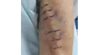
Rupture of AV Fistula
Repeated punctures of the fistula for dialysis with overlying infection lead to the thinning of its walls making it weaker and consequently rupture of the fistula in a few cases.
How is the surgery done if the AV fistula is ruptured?
If ruptured, emergency surgical management is required to control the blood loss and either ligate the fistula or repair it and form a new AV Fistula at the same time.
This is done by microvascular surgery done under a microscope. After repair, extra care is taken to confirm the patency of the fistula and to maintain the blood flow.
Stenosis/ blockage of AVF
Significant stenosis of the vessel lumen is defined as a reduction of more than 50%.
Clinical suspicion of stenosis is confirmed by the presence of several factors:
- reduced quality of dialysis,
- prolonged bleeding after AVF puncture,
- pain in the area of the fistula or
- increased venous pressure.
What is the cause of blockage of the AV fistula?
T hrombosis is a crucial cause of loss of function of an AVF. It usually occurs near the area of anastomosis or vein of the fistula.
- High urea in blood (Usually seen in patients with kidney diseases) increases levels of homocysteine or endogenous inhibitors of NO synthase, which could be directly toxic to the inner lining of the blood vessels.
- Vein wall distensibility is controlled by collagen, elastin, and smooth muscle. There is an accumulation of collagen fibers which replace smooth muscle cells in the pre-access cephalic veins, causing a decrease in the elasticity of the vein wall. This process reduces the distensibility of the veins and thus interferes with the proper maturation of the AVF.
- Stenosis may also be due to compression by an abscess or hematoma.
How is blockage of AV fistula diagnosed?
- Stenosis is diagnosed by measuring the peak systolic velocity in Doppler ultrasound . Values greater than 400 cm/s indicate the presence of stenosis; a monthly decline in flow by 20-25% is also considered significant for this type of complication.
- Angiography is a reliable technique for determining stenosis but is more expensive and technically demanding.
What is the treatment done for stenosis of AVF?
Treatment involves-
- balloon dilatation of the stenosis,
- stent implantation or
- surgical revision.
Percutaneous transluminal angioplasty (PTA) in the treatment of AV Fistula stenosis improves fistula function and prolongs fistula survival in patients with shorter lesions (<1 cm), but restenosis remains the major problem.
Heart failure
Heart failure in patients with AV Fistula occurs only in individuals with previously chronic heart disease. Preliminary data show a trend towards left ventricular hypertrophy (LVH) in patients with an AVF.
How is heart failure treated in patients with AV fistula?
Heart failure is a medical emergency and is treated in an intensive care setting. Medications to strengthen the pumping action of the heart are given and to decrease the burden of work of blood pumping over the heart muscles is tried to reduce by giving medical treatment.
AV fistula-Induced Ischemia (Steal Syndrome)
What is steal syndrome?
Reduced blood flow distal to the AV Fistula, leads to decreased oxygenation, ischemia, and gangrene of the hand in a few cases.
What is steal syndrome in patients with AV fistula?
In most cases, an AV Fistula does not compromise perfusion of the hand but can happen in a few elderly patients and diabetics.
What are the symptoms of steal syndrome in patients with AV fistula?
- Reduced movement of the wrist with a cold hand and
- A color change to pale yellow, or purple
- Pain at rest and during exercise and
- Blackening of the fingers.
What is the incidence of AV fistula-induced ischemia in patients?
There is evidence that the steal syndrome in risk groups may occur in 75-90% of patients after the creation of an AVF. This phenomenon remains clinically without symptoms until the moment when compensatory mechanisms for perfusion by peripheral arteries are exhausted.
How is steal syndrome treated in patients with AV fistula?
Treatment of this condition is difficult and the risk of amputation of fingers and the forearm is great.
Can steal syndrome be prevented in patients?
Attention must be focused on prevention, which includes-
- adequate preoperative assessment, use of Doppler ultrasound, and
- a precise surgical technique that involves arteriotomy no greater than 7 mm as well as being within the range of a 90-180° angle of anastomosis .
Failure to use these precise surgical techniques may lead to increased resistance and reduced blood flow.
Ischemic Polyneuropathy-AV Fistula
Ischemic neuropathy is most common in diabetic patients, especially when the brachial artery is used for the creation of vascular access.
It is manifested by weak forearms, in the immediate preoperative period (typically within hours), severe pain, and altered sensations.
Neurological examination shows weakness in distal muscle groups and decreased sensations in the area of the median nerve. The frequency of such complications varies between 1 and 10%.
Lymphedema-AVF
This is a condition in which there is swelling of the whole limb below the AV fistula caused by blockage of lymph-draining vessels from the limb.
Treatment is usually conservative in mild cases where compression bandages, limb elevation, and rest are advised.